2023 Rising Stars in Reproductive Biology Webinar Series
Program:
Illuminating the (uterine) path: from embryo movement to implantation
Although much is known about the molecular signaling during implantation, the uterine 3D architecture that facilitates embryo development remains unknown. Imaging the mouse embryo and the uterine milieu simultaneously we uncovered patterns of embryo movement and dynamic shape changes in the uterine lumen and glands in preparation for implantation. When applied to mouse mutants with known implantation defects, this method detected striking peri-implantation abnormalities in uterine morphology that cannot be visualized by histology. Analyzing the uterine and embryo structure in 3D for genetic mutants, hormonal perturbations and pregnancies treated with pathway inhibitors is helping us uncover novel molecular pathways and global structural changes that contribute to successful implantation of an embryo. Our studies have implications for understanding how structure-based embryo-uterine communication is key to determining an optimal implantation site, which is necessary for the success of a pregnancy.
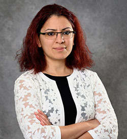
Speaker: Dr. Ripla Arora, Assistant Professor, Department of Obstetrics, Michigan State University

Sex-differences in immune aging: are we missing half of the picture?
Neutrophils are the most abundant human white blood cell and constitute a first line of defense in the innate immune response. Neutrophils are short-lived cells, and thus the impact of organismal aging on neutrophil biology, especially as a function of biological sex, remains poorly understood. We have generated a multi-omic resource of mouse primary bone marrow neutrophil from young and old female and male mice, at the transcriptomic, metabolomic and lipidomic levels. We identified widespread regulation of neutrophil ‘omics’ landscapes with organismal aging and biological sex. In addition, we leveraged this data to predict functional differences, including changes in neutrophil responses to activation signals. To date, this dataset represents the largest multi-omics resource for neutrophils across sex and ages. This resource identifies neutrophil characteristics which could be targeted to improve immune responses as a function of sex and/or age.
Speaker: Dr. Bérénice Benayoun, Assistant Professor of Gerontology, USC

Bérénice Benayoun, Ph.D., is an Assistant Professor of Gerontology at the USC Leonard Davis School of Gerontology. Benayoun’s PhD work focused on a transcription factor whose mutations lead to a human syndrome associated to premature menopause. During her post-doctoral training, she identified a chromatin signature of cell identity remodeled with aging, raising questions about the lifelong stability of cellular identity. Her lab’s research focuses on age-related ‘omic’ changes, and how they interact with sex to shape aging. Her lab is also pioneering the naturally short-lived African turquoise killifish Nothobranchius furzeri as a new vertebrate model for aging research.

